Tissue
Class- IX
Plant tissues: Plant tissues are classified into
two types:
·
Meristematic tissues
·
Permanent tissues
Meristematic tissues: it keeps on dividing
Features of meristematic tissues:
- Cells of these tissues have thin elastic walls, made up of cellulose
- Cytoplasm is dense with nucleus present in the centre of the cell
- No vacuole or very few vacuoles are present
- Cells are completely arranges without interference of spaces
- It is further divide into three parts:
1.
Apical meristemtic:
·
Present at the root and shoot tips.
·
Helps in longitudinal growth of plant
2.
Intercalary meristematic:
·
Present at nodes (just above or below the node)
·
Help in longitudinal growth by increasing
intermodal length
3.
Lateral meristematic:
·
Present parallel to the length of the plant. It
is present just below bark.
·
Helps in increasing girth of the plant.

Permanent
tissues:
- It is formed by meristematic tissue
- Meristematic tissues undergoe differentiation to form permanent tissue
- Cell differentiation is a process by which cells acquire permanent shape and size to perform one special function.
- Cells of permanent tissues do not divide further.
- Permanent tissues can be classified as:
·
Simple permanent tissue
·
Complex permanent tissue
Simple
permanent tissue: (consists only one type of cells)
It
is of following types:
1.
Parenchyma
2.
Collenchyma
3.
Sclerenchyma
1.
Parenchyma:
- It is basic permanent tissues made up of unspecialized cells.
- Cell wall is thin and made up of cellulose.
- Cell has large central vacuole and dense cytoplasm.
- Cells are closely packed and have inter cellular spaces.
- It is most abundant tissues and is found as packaging tissue in all plant organs such as fruit, root and seed.
- It acts as a storage tissue when present in stem and roots ( stores water and minerals)
- In leaf parenchyma cells contain chloroplast and help in photosynthesis. Such parenchyma is call Chlorenchyma.
- In aquatic plants, parenchyma cells are loosely arranged with large intercellular spaces. Such parenchyma is called Aerenchyma. It helps plant to float.
2.
Collenchyma: (made up of living cells)
- It has a cell structure similar to parenchyma tissue but cell wall has thickenings of pectin. These thickenings are more prominent at the corners that are why no intercellular spaces exist between the cells.
Location:
It is found just below epidermis in
stem and leaves of dicot (absent in roots).
It provides flexibility to these organs so that plant can stand against
strong winds. It allows plant to bend without breaking.
It also provides mechanical strength.
3. Sclerenchyma
: (made up of dead cells)
- Cells are long and narrow.
- Cells have heavy deposits of lignin on their walls. Thickenings are so thick that there is no space inside the cell.
- Lignin act like cement and harden these cell.
Location: Present in stem, in veins,
around vascular bundles, covering of seeds and nuts, pulp of fruits (guava,
apple), and husk of coconut.
Function: provide mechanical strength.

Protective
tissues:
- It provide protection to plant it is combination of simple permanent tissue, specially for protection of plant.
- Two type of protective tissues are formed:
1 Epidermal tissue:
- It exists in the form of outer most layer in all plant organs called epidermis
- Epidermis is a single layer in most of the plants byt in xerophytes (desert plants )it can be made up of many layers
- Cells have structure similar to parenchyma nut cell walls are thicker. Cell is thicker in outer surface lateral walls than inner side.
Function:
- It is a protective layer and forms a continuous layer (no inter cellular spaces)
- In lower leaf epidermis small pores are present which help in the exchange of gasses of carbon dioxide and oxygen and transpiration. Stomata are guard by bean shaped guard cells.
- Cells of epidermis tissue produce waxy coating called cuticle. Cuticle is thick xerophytes and prevents loss of water.
- Epidermises of roots produce root hairs which help in absorption of water and minerals by increasing surface area for absorption.
- Cuticle
also provides protection against mechanical injury is parasitic infections.
2.
Cork tissue:
- In old stems and roots epidermis tissue is replaced by a strip of meristematic tissue. This forms thick layers of barks (cork) of the tree.
- Cells of cork are dead and compactly arranged without intercellular spaces. They have deposits of suberin as these cell walls make cork, impervious to form gases and water.
Complex permanent tissue
- This tissue is made up of different types of cells which co-ordinate with each other to perform a common function.
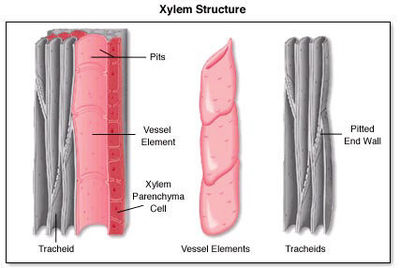
- Xylem and phloem (vascular tissue) are complex permanent tissue.
- Both these tissues are present together in the form of patches called vascular tissues.
- They are conducting tissues are a distinguish feature of terrestrial plants.
Xylem: (conducts water and minerals)
- It consist of following component
·
Vessels
·
Tracheids
·
Xylem parenchyma
·
Xylem sclerenchyma
- Tracheids and vessels are main conducting elements and water and minerals flow through them vertically upwards.
- Xylem parenchyma stores food and also helps in vertically conduction.
- Xylem fibers are supportive in function.

Phloem:(conducts food, hormones, amino acids, etc)
It
consists of following elements:
·
Sieve tubes
·
companion
cells
·
Phloem parenchyma
·
Phloem sclerenchyma
All phloem elements are
living except sclerenchyma.
·
Sieve tubes are tabular cells with sieve like
perforated ends called sieve plate. When mature sieve tubes do not have
nucleus.
· Companion cells are living cells they are
associated with sieve tubes and control their functioning.
· Phloem parenchyma stores food and phloem
sclerenchyma is supportive in function.
· Conduction in phloem takes in both upward and
downward direction in the leaf.

Animal tissues:
The animal tissues have been classified
into four major types depending on the functions they perform.
1.
Epithelial tissue
Structure:
I.
The cell are closely packed and are without
inter cellular spaces.
II.
The lower most layers of the cells rests on a
non cellular basement membrane composed of collangenous fibers.
III.
The free surface of cells may be modified into
cilia and microvilli.
IV.
Epithelial tissue could be simple i.e. made up
of single layer so cells are compound i.e. made up of many layers of cells.
Location:
Epithelial tissue forms a continuous layer all over the external
surface of the body, the skin surface layers of mouth, alimentary canal and
lungs are made up of epithelial tissue.
Function:
1. Protection:
epithelial tissue covers the entire body surface; thereby it protects the
underlying cells from drying, injury, bacteria or viral infections.
2. Exchange
of material being: extremely thin, simple epithelium allows diffusion of gases
or materials.
3. Absorption:
cells help in absorption of water and other nutrients as in intestine
4. Elimination
of water products: epithelial tissue like those in nephron as sweat glands
helps in removal of waste from the body.
5. Secretion:
number of epithelial cells is modified to produce secretion which could be in the
form of mucous, enzymes or hormones.
Epithelial tissue can
be classified into:
|
Type
|
Structure
|
Location
|
Function
|
|
Squamous
epithelium
|
Thin
flattened cells with a centrally placed nucleus. Irregular shaped cells,
compactly arranged.
|
Form
lining of mouth, oesophagus, and lungs.
Inner
lining of blood vessels
Cover
the skin surface.
|
Diffusion
of material or exchange of gases
Protection
from chemical injury entry of germs or from drying.
|
|
Cuboidal
epithelium
|
Cube
like cells with a central, spherical nucleus. Appear hexagonal in surface
view.
|
Parts
of nephron, lining of salivary pancreatic and sweat ducts.
|
Secretion,
excretion and absorption.
|
|
Columnar
epithelium
|
Tall
pillar or column like cells with nucleus at the base. Generally have mucus
cells in between.
|
Lining
of stomach, intestine, and gall bladder.
|
Secretion,
excretion and absorption. Mucous lubricates the passage.
|
|
Ciliated
epithelium
|
Certain
cuboidal and columnar epithelium have cilia at their free ends, cilia are
thin, hair like projections that move to and fro.
|
Oviducts,
trachea bronchioles and in parts of nephron in kidney.
|
Movement
of cilia directs the flow of fluids in a particular one direction.
|
|
Glandular
epithelium
|
Cuboidal
and columnar epithelium and are modified into glands.
|
Salivary,
gastric, intestinal, sweat gland adrenal thyroid glands.
|
Secrete
enzymes, mucous or hormones
|

Nervous tissue:
Nervous tissue is made up of millions of nerve
cells called neurons. The neurons are highly specialized cells. Brain, spinal
cord, and nerves are all composed of neurons.
Structure of a nerve cell or neuron:
1. A
neuron consists of two distinct parts: i) cell body or soma ii) cytoplasmic
processes.
2. The
cell body contains the nucleus and granule cytoplasm.
3. From
the cell body extent out 2 kinds of cytoplasmic processes called dendrite and
axon.
4. Dendrites
are small short, fine branched process, numerous in number impulses towards the
cell body.
5. Axon
is a long cylindrical generally unbranched single process and ends in many
branches called terminal end fibers. Axon conducts the nerve impulses away from
cell body.
6. The
axon may be covered by a fatty myelin sheath.
Connective tissue
Structure: A
composite tissue, it has following three basic components:
1.
Cells- living part, loosely spaced, embedded in
the matrix
2.
Fibers- non living part scattered in between the
cell.
3.
Matrix- basic ground tissue may be jelly like
fluid or dense. Matrix decides the nature and function of a connective tissue.
|
|
Have soft matrix.
Cells are closely packed.
|
Fill the space inside organs like packaging
tissue.
Support internal organs.
Helps in repair of tissues.
|
Between skin and muscles.
Around blood vessels and nerves.
In the bone marrow
|
|
Adipose
|
Has soft matrix.
Cells are closely packed.
Cells are filled with fat globules.
|
Stores fats and acts as an insulator.
Acts as a source of energy reserve.
|
Below the skin.
Between internal organ like kidney, heart,
eyes, etc.
|
|
Ligament
|
Has soft the little matrix.
Cells are densely packed.
Loose network of yellow fibres.
|
Connects two bones at the joint.
Highly elastic and considerable strength.
|
|
|
Tendons
|
Have soft little matrixes.
Consist of white collagen fibers
|
Connect muscles to bone.
Tissue has great strength but limited flexibility.
|
|
|
Bone
|
Matrix is solid and hard and made up of
calcium and potassium salts.
Bone cells called osteocytes are arranged
around harvesian canal.
Harvesian canal has blood vessel and nerve
fibers.
|
Forms the frame work/ skeleton of the body.
Anchors the muscles.
Support and protects main organs of the
body.
|
Skeleton
|
|
Cartilage
|
Solid matrix composed of protein and sugars.
Widely spaced cells called chondrocytes.
Hard but flexible tissue
|
Smoothens bone surfaces at joint.
Forms some parts of skeleton
|
Present at the ends of bones.
At the tip of nose.
Ear penna
Trachea
Larynx
|
|
Blood
Rbc
Wbc
Platelets
|
Fluid matrix called blood plasma.
Blood cells called corpuscles float in
matrix.
They are of three types
1.
RBC(erthyocytes)
2.
WBC(leucocytes)
3.
Platelets (thromocytes)
|
RBC transport O2 and CO2 .
WBC protects against diseases.
Platelets help in blood clotting.
Transport food hormones waste materials.
|
|

Muscular tissue:
Features-
1. Muscular
tissue consists of elongated cells called muscle fibers.
2. This
tissue has the capability to contract and relax causing movement due to
presence of contractile proteins (actin, myosin).
Types of
muscular tissue: 3 types of muscle tissue
|
Feature
|
Striated
|
Unstraited
|
Cardiac
|
|
Structure cell
shape
|
Elongated , cyndrical,
unbrached
|
Spindle shaped with
tapering ends
|
Elongated,
cylindrical branched
|
|
Nucleus
|
Multinucleated with
peripheral nucleus
|
Uninucleated with
centrally located nucleus
|
|
|
Striations(bands)
|
Has alternate light
and dark striations
|
No striations
|
Faint regular
striations
|
|
Interrelated dics
|
Absent
|
Absent
|
Present
|
|
Function
|
Voluntary muscles
Undergo rapid
contraction, tired easily
|
Involuntary muscles
Undergoes slow and
rhythmic contraction, do not get tired easily
|
Involuntary muscles
Undergo continuous
and rhythmic contractions and relaxation, without getting fatigue
|
|
Location
|
Called skeletal
muscles, attached to bones
|
Called smooth
muscles, present in organs like ureters stomach , intestine
|
In walls of heart
|

No comments:
Post a Comment